Exploring Cells, Microscopy and Transport Mechanisms in GCSE Biology
This work has been verified by our teacher: 20.02.2026 at 11:44
Homework type: Essay
Added: 17.02.2026 at 15:28
Summary:
Discover key GCSE Biology concepts on cells, microscopy, and transport mechanisms. Build a strong foundation for exams with clear, detailed explanations.
Understanding Core Biological Concepts: Cells, Microscopy and Transport Mechanisms in Living Organisms
Biology, underpinning much of modern science and medicine, begins with the tiny structures that make up all living things: cells. At the heart of GCSE Biology, grasping the core principles surrounding cellular structure and function, the revolutionary use of microscopes, the astonishing range of cell specialisations, and the movement of substances within and between cells, forms the bedrock of further scientific understanding. The ability to look inside the living world, to decipher its subtleties and adaptations, not only furthers knowledge but empowers future studies in health, agriculture, and technological innovation. This essay will explore the importance of microscopes in unveiling the secret lives of cells, will detail the differences and commonalities among animal, plant, and microbial cells, and will discuss the means by which cells transport essential materials. These fundamental topics form a coherent foundation, setting the stage for life sciences across the United Kingdom’s educational curriculum.
---
1. The Role and Evolution of Microscopy in Biological Studies
1.1 Historical Context and Importance
Long before the development of microscopes, naturalists such as Robert Hooke and Antonie van Leeuwenhoek peered through primitive lenses, opening up realms no human eye had previously witnessed. Hooke’s seminal observations in the 17th century, capturing the honeycomb appearance of cork, led him to coin the term "cell"—a moment that reverberated through scientific history. The subsequent refinement of optical instruments transformed biology from educated guesswork, long intertwined with speculative observation, into a rigorous experimental science. In the United Kingdom especially, with scientific societies such as The Royal Society propelling curiosity and collaboration, microscopy soon became an indispensable tool.1.2 Types of Microscopes and Their Capabilities
Light Microscopes, using beams of visible light and glass lenses, remain a staple in schools and teaching laboratories due to their simplicity and safety. Capable of magnifying samples up to around ×2000, light microscopes allow the observation of cells, many organelles, and even the dynamic movement within living cells themselves. However, their resolution is inherently limited by the wavelength of light, usually not finer than 200 nanometres.Electron Microscopes revolutionised cell biology in the 20th century. By employing beams of electrons—much smaller than photons—these instruments have pierced the barrier of visible light and magnified specimens up to two million times, with resolving capabilities measured at just 0.2 nanometres. This leap has enabled biologists to study the ultrastructure of organelles; for instance, the intricate folds of mitochondria or the detailed anatomy of chloroplasts. Transmission Electron Microscopes (TEM) produce highly detailed cross-sectional images by passing electrons through thin slices, whilst Scanning Electron Microscopes (SEM) render three-dimensional surfaces, invaluable for examining bacteria or pollen grains.
1.3 Components and Functions of a Light Microscope
A typical light microscope comprises several interdependent parts: the stage, on which the slide sits; the mirror or light source, illuminating the sample; objective lenses of varying magnification; the eyepiece lens for secondary magnification; and focusing knobs (coarse and fine) to clarify images. Each part is essential for optimising visibility. For instance, improper slide placement or inadequate focusing often results in blurred or distorted images. When preparing slides, care must be taken to use a coverslip and avoid trapping air bubbles, while stains such as iodine or methylene blue can greatly enhance contrast, revealing nuclear and cytoplasmic features with clarity.1.4 Measuring Magnification and Resolution
The formula: _magnification = image size / actual size_ is a frequent companion for biology students. For example, if a drawn image of a leaf cell measures 40 mm across, but the actual cell is only 40 μm wide, the magnification is 40,000 μm / 40 μm = ×1000. However, without adequate resolving power—the minimum distance at which two points can be distinguished as separate—greater magnification just produces a bigger blur. Recognising the value of fine resolution explains why, when studying organelles like ribosomes or the detailed structure of cell membranes, electron microscopes are indispensable.---
2. Cellular Structure: Comparing Animal, Plant and Microbial Cells
2.1 Introduction to Cell Theory and Cell Diversity
The assertion, central to cell theory, that every living thing consists of cells, forms the unshakeable cornerstone of all biology. Yet this deceptively simple idea unfolds into stunning diversity: from the humblest bacterium, invisible to the naked eye, to the towering beech trees dotting British woodlands, all are underpinned by cells tailored to specific environments and functions.2.2 Anatomy and Functions of Animal Cells
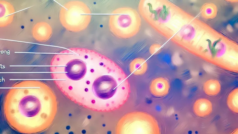
A typical animal cell, as found in the lining of a human cheek, contains a set of core organelles. The nucleus acts as the command centre, housing genetic instructions on chromosomes and wrapped in a protective double membrane. The surrounding cytoplasm is a watery matrix where enzymes orchestrate the multitude of life’s chemical reactions. Dotted throughout are mitochondria, the so-called “powerhouses”, extracting energy from nutrients through aerobic respiration to supply adenosine triphosphate (ATP). Ribosomes, found either free in the cytoplasm or attached to the endoplasmic reticulum, translate genetic instructions into proteins. All is encased by the cell membrane, a semi-permeable barrier that not only keeps cellular contents safe but also carefully regulates the exchange of substances, mediating both defence and communication.2.3 Anatomy and Functions of Plant Cells
Plant cells share many features with animal cells but display three major differences, crucial for their survival and role in ecosystems. The cell wall, predominantly made of cellulose, encases each cell, lending strength and rigidity and aiding plants in standing upright. Chloroplasts, unique to plants, contain the green pigment chlorophyll and are the site for photosynthesis, which drives the conversion of light energy to glucose—sustaining not only plants but, indirectly, all animal life in Britain’s food webs. The roomy permanent vacuole stores dissolved sugars and maintains internal pressure (turgor), preventing wilting. While mitochondria, ribosomes, nuclei, and the cytoplasm are present as well, plant cell features highlight adaptations for stationary, sunlight-dependent life.2.4 Prokaryotic Cells and Their Distinctiveness
Prokaryotic cells, especially bacteria, are simpler yet remarkably versatile. Instead of a membrane-bound nucleus, their DNA exists as a single circular loop, sometimes accompanied by small rings called plasmids—crucial in the rapid spread of antibiotic resistance in pathogens like *Staphylococcus aureus* in UK hospitals. Certain bacteria bear a slime capsule for extra protection or develop flagella, whip-like tails for locomotion. Despite their diminutive size, bacterial cells underpin essential processes, from recycling nutrients in a compost heap to nitrogen-fixing in pea plant root nodules near farms in East Anglia.---
3. Cellular Specialisation: How Cells Adapt to Specific Functions
3.1 The Process of Cell Differentiation
Every multicellular organism develops from a single cell—a fertilised egg—which then divides and differentiates into an array of specialised cells, each tailored for a dedicated function. This transformation is dictated partly by gene expression and partly by signals from neighbouring cells. In the developing embryo, genes switch on or off to direct the cell to become, for instance, a red blood cell or a neurone. Such specialisation continues throughout the lifetime, as seen in skin healing or new root growth in plants.3.2 Specialised Animal Cells
Some examples illuminate the process. Nerve cells (neurones) are elongated, with extended axons and branched dendrites, designed to transmit electrical impulses rapidly over long distances—key for human reflexes. An insulating myelin sheath speeds up this conduction, essential for activities ranging from playing cricket to learning a new tune on the piano. Muscle cells teem with mitochondria, reflecting their high energy demands during contraction, while their protein filaments slide past each other to generate movement. Sperm cells, equipped with streamlined heads, enzyme-filled acrosomes, and powerful flagella, are adapted for swimming towards and fertilising the egg—central to sexual reproduction.3.3 Specialised Plant Cells
Root hair cells extend finger-like projections into the soil, maximising surface area for water and mineral uptake; their cytoplasm contains many mitochondria to fuel active transport against concentration gradients. Palisade cells in leaves, densely packed with chloroplasts, capture sunlight efficiently. Xylem vessels, reinforced by lignin and dead at maturity, form sturdy pipes for water transport from root to leaf—a process visible in the rise of sap in British oak trees each spring. In parallel, phloem cells, with perforated sieve plates and companion cells, distribute sugars made during photosynthesis to growing tissues and storage organs.3.4 Integration into Tissues, Organs, and Systems
Specialised cells seldom act alone. Groups integrate into tissues (like muscle in the arm or vascular tissue in a daffodil stem), combine into organs (such as the stomach or a sycamore leaf), and ultimately function as coordinated systems—the circulatory, nervous, or transport systems—working in harmony to maintain the balance (homeostasis) necessary for survival.---
4. Mechanisms of Particle Movement: Diffusion and Osmosis
4.1 Diffusion: Principles and Biological Significance
Diffusion is the spontaneous movement of particles from an area of higher to lower concentration, powered by the constant random motion of molecules. Factors such as a steeper concentration gradient, higher temperature, and greater surface area increase diffusion’s rate—a vital principle in understanding why alveoli in human lungs are so thin-walled and numerous, enabling rapid oxygen uptake and carbon dioxide removal with every breath. Similarly, inside the cell, oxygen moves in and carbon dioxide out by diffusion, supporting respiration.4.2 Osmosis: Specialised Movement of Water
Osmosis, a special case of diffusion, involves the movement of water molecules across a partially permeable membrane from less concentrated (more watery) to more concentrated solutions. In plant roots, osmosis draws water from soil into cells, maintaining turgor that keeps leaves rigid. A familiar school experiment uses potato chips immersed in sugar solutions of varied strength; when placed in dilute solution, the potato swells as water enters, but in concentrated solution, it shrinks as water leaves—clear, hands-on evidence of osmosis at work. In animal cells, the stakes are higher: too much water gained and red blood cells burst (lysis); too little and they shrivel (crenation).4.3 Comparing and Contrasting Diffusion and Osmosis
Although both processes move substances down concentration gradients and require no energy, osmosis is unique to water and needs a selectively permeable membrane, whereas diffusion involves a wider variety of gases and solutes, sometimes not needing a membrane at all. Together, they facilitate nutrient uptake, waste removal, and the balance of fluids vital to life.---
Conclusion
The microscopic universe, once hidden from view, is now central to our understanding of biology thanks to the development of microscopy. Mastery of cell structure and specialisation illuminates the incredible adaptability and function of life’s building blocks, from bacteria in our gut to the leaves overhead. The movement of substances by diffusion and osmosis connects all these processes, underpinning the most essential tasks of survival. Each topic, though distinct, forms a tightly woven net of concepts that support not just scientific knowledge but practical progress in medicine, farming, and beyond. For every GCSE biology student, embracing both theory and hands-on investigation is the key to unlocking the wonders tucked away beneath the microscope lens and within each living cell.---
Additional Suggestions for Further Study and Revision
To deepen understanding and prepare for examinations, students are encouraged to:- Practise using school microscopes and develop skills in preparing slides, observing and drawing what is seen. - Construct models of plant, animal, and bacterial cells using household materials to reinforce the position and function of organelles. - Conduct simple diffusion experiments, such as observing the spread of food colouring in water or timing the movement of salt through agar jelly. - Set up and interpret osmosis experiments with potato strips, weighing changes in different solutions. - Draw annotated diagrams and construct flow charts to chart the journey from single cell to complex organism and the vital movements that sustain life.
In doing so, the vivid processes underpinning all living things become tangible, setting the stage for future scientific discovery.
Rate:
Log in to rate the work.
Log in