Explore the human muscular system’s structure and function to understand muscle types, movement mechanics, and their role in daily life and biology studies.
Comprehensive Exploration of the Human Muscular System: Structure, Function, and Mechanics
The human muscular system is a marvel of biological engineering, underpinning nearly all facets of movement, posture, and physical interaction with the world. Without the seamless operation of muscles, activities as diverse as sprinting for a bus, maintaining an upright stance during a formal assembly at school, or even the subtle facial expressions exchanged across the dinner table, would be impossible. The interplay between the muscular and skeletal systems ensures not only mobility but also the stabilisation necessary for daily life. This essay will delve into the key themes of the muscular system: classifying the different muscle types, delving into skeletal muscle structure from the macroscopic to the microscopic, clarifying the molecular choreography behind contraction, scrutinising the role of joints and levers in movement, and highlighting the clinical and practical significance of muscular knowledge in the United Kingdom context.
Classification and Types of Muscle Tissue
In the human body, muscular tissue is divided into three main categories, each adapted to its role. Skeletal muscle is the most familiar—these are the ‘muscles’ most people envision, lying attached to the bones via strong tendons. They are characterised by their striated appearance under the microscope, their multinucleated cells, and, crucially, their voluntariness; conscious thought allows us to command these muscles, whether lifting a copy of Shakespeare or typing a biology essay. Cardiac muscle, exclusive to the heart, shares skeletal muscle’s striations but is involuntary, controlled by the body’s intrinsic electrical signals rather than conscious effort—a pulse never depends on our willpower. Smooth muscle, found lining the walls of internal organs like the intestines and blood vessels, lacks striations and governs functions such as digestion and blood flow without conscious intervention. For the purposes of understanding movement as it relates to A-Level and IB Biology, skeletal muscle is paramount, given its role in voluntary locomotion and its unique structural features.
Organisation of the Skeletal Muscular System
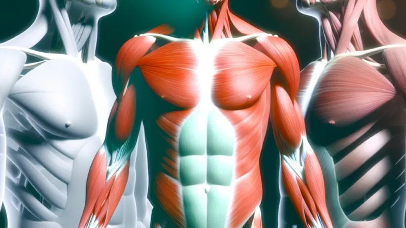
On a grand scale, skeletal muscles operate in groups attached across joints, arranged in antagonistic pairs; the classic example is the biceps and triceps at the upper arm, whose coordinated action allows precise control over flexion and extension at the elbow joint. Each muscle attaches to bone via a tendon—a tough, fibrous band enabling efficient transfer of force. Cutting into the structure, each muscle is comprised of bundles called fascicles, which are themselves bundles of individual muscle fibres, or cells. These muscle fibres are notable for their length and multinucleation, encompassed by the sarcolemma (cell membrane). Within every fibre lies a dense packing of myofibrils, which are cylindrical arrays of contractile proteins. The fibres, fascicles, and entire muscles are ensconced within layers of connective tissue—endomysium surrounds fibres, perimysium encases fascicles, and epimysium wraps the whole muscle—providing insulation, support, and a conduit for nerves and blood vessels. Tucked within each fibre, the sarcoplasmic reticulum sequesters calcium ions critical for contraction, while mitochondria supply the ATP energy underpinning muscle activity. This hierarchical arrangement enables both strength and finesse, allowing for precise control over a vast range of movements, from the sweep of a cricket bat to the steady pressure of a surgeon's hand.
The Sarcomere: The Functional Unit of Contraction
At the heart of muscular function lies the sarcomere—the fundamental contractile unit within each myofibril. Sarcomeres are bordered by structures known as Z lines and are packed in series throughout the length of each myofibril. Their highly ordered arrangement leads to the characteristic striped ("striated") appearance observable under light microscopy—a feature first documented in 19th-century British anatomical studies. Within each sarcomere, interlacing thick filaments (composed primarily of myosin) and thin filaments (mainly actin) overlap in a specific pattern. Actin filaments are anchored at the Z lines and present binding sites, while myosin filaments, with their protruding “heads”, lie centrally, ready to engage these sites. Accessory proteins, namely tropomyosin and troponin, drape along actin filaments, serving as gatekeepers that regulate access to the myosin-binding sites, ensuring contraction is a tightly controlled event.
The Molecular Mechanism of Muscle Contraction
The sliding filament theory, developed through decades of physiological research partly spearheaded by British scientists such as Sir Andrew Huxley, describes how muscles contract at the molecular level. When a voluntary movement is initiated, a motor neuron releases the neurotransmitter acetylcholine at the neuromuscular junction, generating an electrical current that traverses the sarcolemma and dives into the fibre via T-tubules. This signal triggers the sarcoplasmic reticulum to flood the muscle cell with calcium ions. Calcium binds to the troponin complexes on the actin filaments, shifting the position of tropomyosin and exposing the binding sites for myosin.
The myosin heads attach to actin, forming cross-bridges, and execute a power stroke, pulling the actin filaments inward and thereby shortening the sarcomere—a process repeated thousands of times across the muscle fibre. ATP molecules bind to the myosin heads, causing them to detach from actin; hydrolysis of ATP then recocks the heads, priming them for the next cycle of attachment and pulling. This rapid, cyclical event underpins all voluntary movement, from the deft fingerwork of a pianist preparing for a Grade 8 ABRSM performance to the explosive push of a rugby forward. When neural stimulation ceases, calcium is actively pumped back into the sarcoplasmic reticulum. Without calcium, troponin and tropomyosin revert, masking the binding sites and allowing the muscle to relax and lengthen back to its original state.
Skeletal Joints: Structures Facilitating Movement
Muscles alone cannot generate movement without their close relationship with the skeletal system, particularly the joints, or articulations, which serve as pivot points for motion. Synovial joints, such as those found at the knee, elbow, shoulder, and hip, are the most complex and mobile in the human body. Their structure, featuring articular cartilage, a synovial fluid-filled cavity, and a robust joint capsule, is tailored to reduce friction while maintaining mechanical stability. Cartilage ensures the bones glide smoothly, acting as a shock absorber—a feature vital in absorbing impact during activities like running for a school sports day or leaping for a line-out at Twickenham. Synovial fluid lubricates further and nourishes the cartilage, while ligaments entwine the joint capsule, preventing dislocation. The range and type of movement permitted depend on the joint’s architecture: hinge joints (elbow, knee) allow flexion and extension; pivot joints (between the atlas and axis vertebrae) enable rotational movement; and ball-and-socket joints (shoulder, hip) permit movement in multiple axes—a structure that enables the windmill action of a cricket bowler or the elegant extension of a ballet dancer.
The Principle of Levers in the Musculoskeletal System
The interaction between muscles, bones, and joints is often best understood through the principle of levers. In mechanical terms, a lever requires an effort (force), a fulcrum (pivot point), and a load (resistance). In the human body, bones serve as the levers, joints are the fulcrums, and muscles provide the effort. The elbow, for instance, acts predominantly as a third-class lever: when lifting a cup of tea, the biceps provide the effort applied between the fulcrum (elbow joint) and the load (teacup in hand). This arrangement does not maximise force but offers an enormous speed and range of movement, a trade-off that suits most human actions. First- and second-class levers, such as the action of the neck muscles or the rising of the body onto the toes, illustrate how the body adjusts mechanics for differing functional needs.
Coordination of Muscle Groups During Movement
Effective movement depends not on solo muscle action but on collaboration between groups. Muscles typically work in antagonistic pairs: as one, the agonist, contracts, the opposing antagonist relaxes. For instance, during elbow flexion (as when performing a biceps curl), the biceps contracts while the triceps relaxes; for extension, their roles reverse. Synergists act alongside agonists to refine movement, and fixator muscles hold bones steady, like the scapular muscles stabilising the shoulder blade as the arm lifts. This sophisticated organisation allows humans to perform varied and complex activities, from sprinting the 100 metres on Sports Day to writing legibly in exam conditions.
Muscle Fatigue, Repair and Adaptation
Prolonged or intense muscle usage can result in fatigue, often familiar to any sixth-former barely able to climb the stairs after a cross-country run. At root, fatigue stems from depleted ATP reserves, accumulation of lactic acid, and ionic imbalances within the muscle fibre. During rest, the body restores these resources—replenishing calcium stocks, expelling metabolic byproducts, and repairing any microtear damage via satellite cells. Repeated training prompts remarkable adaptations: hypertrophy (increase in muscle fibre size), greater density of capillaries, and a surge in mitochondrial number, all of which underpin the improved endurance and strength seen in trained athletes, professional dancers, or even keen Duke of Edinburgh participants.
Practical Applications and Clinical Relevance
A grounding in the muscular system’s mechanics is invaluable not just in healthcare and biological research, but also for public health, sports performance, and clinical rehabilitation. Conditions such as muscular dystrophy or atrophy highlight the catastrophic consequences of muscle malfunction, while joint diseases like osteoarthritis can severely limit mobility, a growing concern as the UK’s population ages. Physiotherapy relies heavily on knowledge of antagonistic pairs and lever mechanics to devise effective recovery regimes, whether for a sprinter recovering from a hamstring injury or an elderly patient following hip replacement surgery on the NHS. In sports science, understanding how to strengthen both agonists and antagonists is key to injury prevention. Advances in prosthetics and robotics increasingly mimic biological muscle design, borrowing principles from nature’s engineering to improve quality of life for amputees.
Conclusion
From the microscopic interaction between actin and myosin to the sweeping arcs of human athletic achievement, the muscular system epitomises biological complexity and adaptability. Integrating detailed knowledge of muscle structure, physicochemical processes, and biomechanical principles is essential for understanding not only movement but also health, performance, and even technological innovation. Whether in a biology classroom in Manchester, a physiotherapy suite in London, or a research laboratory at Oxford, the study of the muscular system remains central to our understanding of what it means to live, move, and thrive.
Frequently Asked Questions about AI Learning
Answers curated by our team of academic experts
What are the main types of muscles explained in an in-depth essay on the human muscular system?
The main types are skeletal, cardiac, and smooth muscle, each with distinct structures and roles in movement and bodily functions.
How does the structure of skeletal muscle support its function in the human muscular system essay?
Skeletal muscle is made of bundled fibres with myofibrils, enabling voluntary movement and precise control over body actions.
Why is the sarcomere important in the human muscular system structure and function essay?
The sarcomere is the contractile unit in muscle fibres, responsible for muscle contraction and giving skeletal muscle its striated appearance.
What is the role of antagonistic muscle pairs according to the in-depth essay on the human muscular system?
Antagonistic muscle pairs, like the biceps and triceps, work together around joints to control movement direction and coordination.
How do the structure and function of the human muscular system benefit daily life in the UK?
The muscular system enables mobility, posture, and essential tasks, underpinning activities from walking to sports commonly performed in the UK.
Rate:
Log in to rate the work.
Log in