Understanding the Human Skeleton, Joints, Muscles, and Circulatory System
This work has been verified by our teacher: 21.04.2026 at 15:36
Homework type: Essay
Added: 20.04.2026 at 7:39
Summary:
Explore the human skeleton, joints, muscles, and circulatory system to understand their structure, function, and importance for GCSE biology and health studies.
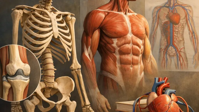
Biology B5 – The Human Skeleton, Joints, Muscles, and Circulatory System: Structure, Function, and Health
The complexity of the human body owes much to the intricate web of systems that maintain our structure and allow life to flourish. Among these, the skeleton, the union of joints and muscles, and the circulatory system are pivotal in shaping who we are. Their interplay forms the foundation of our ability to move, to heal, and to transport vital substances throughout the body. Appreciating the sophistication of these systems is not only a cornerstone of secondary school biology in the United Kingdom, appearing across the GCSE specifications, but also deeply relevant to our own health and wellbeing. By examining their structure, function, and challenges, we develop not only scientific knowledge but an understanding crucial for our everyday lives and future healthcare.
---
The Skeletal System: Framework and Functionality
Purpose and Roles of the Skeleton
Our skeleton is more than just the familiar cranium and long bones often seen in classroom displays; it is an engineering masterpiece, performing diverse and vital roles. Fundamentally, the skeleton provides support, maintaining an erect posture and giving our bodies their characteristic shapes, as observed in the strong, upright frames of footballers or ballet dancers. Equally crucial is protection: the skull secures the delicate brain, while ribs and sternum form a shield around the heart and lungs. The skeleton also enables movement—bones act as levers, and joints as pivots, which muscles pull on to initiate movement, whether that be sprinting in a 100m race or simply raising an arm in class.Its role as a mineral reservoir is less visible but no less significant. Bones store key minerals, particularly calcium and phosphorus, which can be withdrawn into the bloodstream as needed. Most surprisingly, bones are central to blood cell production. Within specialised tissue called bone marrow—abundant inside long bones—new blood cells are created in a process known as haematopoiesis. This keeps us supplied with the oxygen-carrying red blood cells, infection-fighting white cells, and platelets crucial for clotting, all vital to survival.
Types of Skeletons and Evolutionary Adaptations
While all vertebrates share the basic blueprint of an internal skeleton or endoskeleton, invertebrates such as insects rely on external skeletons (exoskeletons). The endoskeleton’s major advantage is its capacity for continuous growth, matching the rate of the growing body—a feature visible as children gain height each year, rather than needing to shed a rigid outer layer like a hermit crab. Furthermore, internal skeletons allow great flexibility and offer multifaceted surfaces for the attachment of powerful muscles. The vertebral column—seen in everything from a herring to the local fox—represents an evolutionary leap, affording both robustness and the potential for a wide range of movement.Bone Structure and Composition
Contrary to the notion of bone as inert matter, it is vibrant, dynamic tissue. Living bone contains specialised cells embedded in a matrix, allowing growth, repair, and remodelling. The shaft of long bones, such as the femur or humerus, is designed for both strength and lightness—its hollow interior reduces weight while still bearing heavy loads. Within many long bones lies the bone marrow, which comes in two types: red, responsible for blood formation, and yellow, which stores fats. The microscopic view of bone reveals a remarkable combination of tough collagen fibres and a hard mineral matrix made mainly of hydroxyapatite, together enabling an optimal blend of rigidity and slight flex, so vital in preventing breaks.Bone Development and Growth
Bone starts as pliable cartilage during foetal development, a feature evident in the small, supple noses and ears of infants. Through ossification, minerals steadily deposit and harden the cartilage into bone. Growth plates, or epiphyseal plates, located near the ends of long bones, are the seats of bone lengthening in children and teenagers. These plates disappear as adulthood approaches, marking the end of height increase. Observing the hands on X-rays is a way doctors can gauge a young person’s "bone age" by assessing the maturity of these plates.---
Cartilage and Its Critical Role in the Skeleton
Cartilage Structure and Properties
Cartilage is a semi-rigid yet flexible tissue found in select regions of the body. It comes in three principal forms: hyaline cartilage, which covers joint surfaces and shapes the nose and windpipe; fibrocartilage, which forms durable pads between vertebrae; and elastic cartilage, which gives the ear its springiness. Essential in the joints, cartilage reduces friction and absorbs shocks—consider how your knees cope with the repeated force of running on the school field.Maintenance and Repair of Cartilage
Unlike bone, cartilage is poorly supplied by blood, making its repair slow and incomplete. As we age, or if we are unlucky enough to damage it (as commonly happens to professional athletes), cartilage thins and deteriorates, raising the risk of joint conditions such as osteoarthritis. This underlines the importance of care and prevention, particularly in sports and high-impact activities.---
Common Skeletal and Cartilage Injuries and Disorders
Fractures and Bone Damage
Bone fractures are common injuries, resulting from accidents, falls, or repeated stress. Young children may suffer greenstick fractures, where the bone bends and partly breaks, much like a fresh twig. In contrast, adults are more prone to complete breaks. Fractures are classified for severity—simple fractures leave skin intact, while compound fractures pierce through, risking infection. The healing process is a finely tuned sequence: an immediate inflammatory response draws repair cells, a soft callous of cartilage forms, which is later replaced by hard bone, eventually remodelling to approximate the original structure.Bone Diseases
Disorders such as osteoporosis emerge later in life, especially amongst post-menopausal women due to decreasing oestrogen levels. Bones become porous and fragile, heightening the risk of hip or wrist fractures from minor falls. Osteoarthritis, in contrast, is a degenerative joint disease caused by the breakdown of cartilage, leading to pain, swelling, and restricted movement. Sadly, cases of osteoporosis and arthritis are well illustrated by stories of elderly relatives or historical figures whose mobility gradually diminished.Precautions and Emergency Response
If there is suspicion of a fracture, especially involving the back or neck, it is crucial to avoid movement—shifting an injured person can cause nerve damage or paralysis. This principle is drilled into St John Ambulance training in British schools, highlighting the importance of proper immobilisation and prompt, professional care.---
Joints and Muscles: The Machinery of Movement
Joint Structure and Types
Joints are marvels of biological engineering. Fixed joints, like those in the skull, fuse bones without movement. Slightly movable joints, such as those joining the vertebrae, balance stability with restricted movement. Freely movable (synovial) joints, including the knee, hip, and shoulder, are designed for a wide range of motion, allowing us to walk, dance, and even play the violin. Each synovial joint consists of bones capped with cartilage, surrounded by an articular capsule, and enclosed in synovial fluid.Ligaments, strong bands of fibrous tissue, stabilise joints while allowing necessary motion. Anyone who has twisted an ankle will appreciate the effectiveness—and vulnerability—of these tough cords.
Roles of Cartilage and Synovial Fluid
The ends of bones are coated with a gleaming layer of articular cartilage, diminishing friction and soaking up the shocks we barely notice. Meanwhile, synovial fluid acts as a biological lubricant, ensuring movement remains smooth even after decades of use—unless disease or injury intervenes.Muscle Attachment and Mechanism of Movement
Skeletal muscles, attached by sturdy tendons to bones, power every voluntary movement. Importantly, muscles can only contract—they cannot push. To facilitate two-way motion at a joint, muscles work in antagonistic pairs: the biceps bends the arm, while the triceps straightens it. This beautifully coordinated dance underlies everything from writing an essay to scoring a goal in a netball match.Joint Injuries and Prosthetics
Joint injuries are common, especially amongst sports enthusiasts, and can result in long-term impairment. Advances in medicine have enabled the replacement of damaged joints with artificial ones, transforming lives—just as National Health Service (NHS) statistics show rising numbers of hip replacements restoring mobility to people who might otherwise face isolation.---
The Circulatory System: Transport and Regulation of the Body
Overview of Circulatory System Structure
The circulatory system comprises the heart, an impressive muscular pump; an extensive network of arteries, veins, and capillaries; and the blood itself. Its multiple roles include the delivery of oxygen from the lungs, absorption of nutrients from digestion, transport of hormones, and export of waste products to related organs.The Double Circulatory System in Humans
Human circulation is double: the heart sends blood on two separate journeys. In the pulmonary circuit, blood travels to the lungs to release carbon dioxide and absorb oxygen. The systemic circuit sends this now-rich blood to every part of the body, powering daily life. This arrangement is more efficient than the single circuit of fish, for example, which explains the higher metabolic capability characteristic of mammals.Heart Anatomy and Function
The human heart is a four-chambered wonder. Blood enters the atria, flows into the muscular ventricles, and is then forced out under pressure. Valves safeguard one-way flow, preventing backwash. The cardiac cycle includes periods of contraction (systole) and relaxation (diastole), processes which can be monitored during routine check-ups with a stethoscope.Blood Pressure and Vascular Differences
Arteries, with their thick, elastic walls, withstand the heart’s forceful output, while thinner-walled veins carry blood back to the heart, helped along by one-way valves, particularly in the legs. Capillaries, just a cell thick, facilitate exchange between the blood and tissues. Blood pressure falls markedly from arteries to capillaries to veins—a principle that underpins measuring blood pressure as a clinical test.Regulation of Heart Rate and Circulation
During exercise or stress, signals from the brain and hormones like adrenaline accelerate the heart rate and increase stroke volume, ensuring that demand for oxygen and nutrients is matched. This ability is why we quickly recover from short bursts of activity or excitement—a benefit highlighted in the rigorous fitness regimes of professional athletes.---
Historical Insights and Scientific Discoveries
Early Understanding of Circulatory Function
Misconceptions abounded for centuries. The Roman physician Galen asserted that blood was produced in the liver, slowly ebbing through the body. It was only in the seventeenth century that William Harvey, a physician educated at Gonville and Caius College, Cambridge, painstakingly demonstrated the heart's true role and the presence of valves. His work, derided at first, ultimately transformed medicine and is celebrated in the corridors of the Royal College of Physicians to this day.Evolution of Knowledge About the Skeleton and Circulation
Microscopy unveiled the living structure of bone, while Victorian X-rays delivered the first anonymous glimpses of internal fractures. More recently, 3D imaging and keyhole surgery have revolutionised diagnosis and treatment—remarkable testimony to the ceaseless progress of science.---
Rate:
Log in to rate the work.
Log in