An In-Depth Essay on the Structure and Role of Cell Organelles
This work has been verified by our teacher: 28.04.2026 at 9:37
Homework type: Essay
Added: 27.04.2026 at 14:02
Summary:
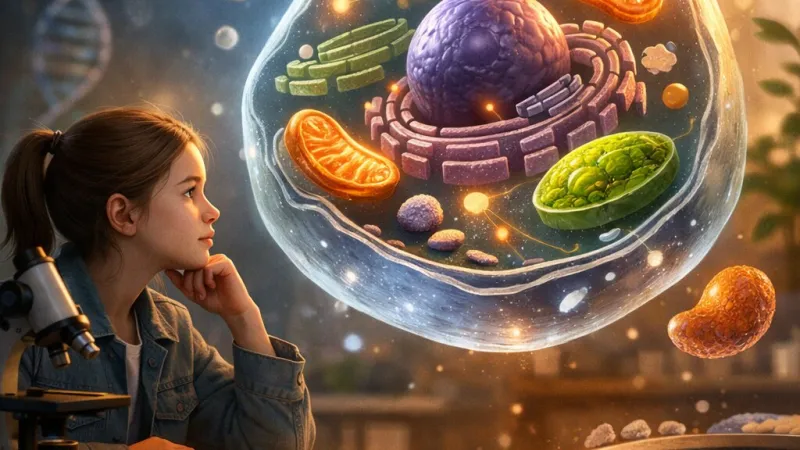
Explore the structure and role of cell organelles to understand how each part supports eukaryotic cell functions and overall organism survival. 🧬
The Marvel of Cell Organelles: Structure, Function, and Their Importance in Eukaryotic Cells
The cell, often celebrated as the “building block of life,” is far from a simple entity. Within its boundaries lie a multitude of specialised compartments known as organelles, each dedicated to ensuring specific aspects of cellular function. This compartmentalisation is not merely a structural peculiarity; it is a brilliant evolutionary solution to the challenge of housing, regulating and coordinating the array of reactions that enable life to persist and adapt. In this essay, the myriad forms and vital functions of cell organelles in eukaryotic cells will be explored; from the command centre of the nucleus to the bustling energy factories found in mitochondria, we will see how these structures underpin not just the survival of individual cells, but the success of complex organisms, including ourselves.
What Are Cell Organelles?
Cell organelles can be succinctly described as distinct subcellular structures, most commonly membrane-bound, that carry out specialised tasks within eukaryotic cells. Unlike prokaryotes—such as bacteria, whose interior is largely an undivided space—eukaryotic cells (those making up plants, animals, fungi, and protists) are distinguished by their intricate internal organisation. Membrane-bound organelles, such as the nucleus or mitochondria, enable the precise separation of biochemical pathways, preventing potentially conflicting reactions from interfering with one another. This level of organisation is absolutely crucial for the advanced metabolism and regulatory complexity exhibited by plants and animals, and is a defining feature separating eukaryotes from their simpler prokaryotic counterparts.The Nucleus: Director of Cellular Affairs
Dominating most eukaryotic cells, the nucleus is easily the most recognisable organelle. Shielded by a double-layered nuclear envelope, which is studded with protein-lined pores, the nucleus keeps the cell’s precious genetic material safe. The envelope’s pores are not passive holes; rather, they carefully regulate the ingress and egress of molecules, selective about what information, energy, and materials traverse in or out. Within the nucleus can be found chromatin (DNA wrapped around proteins called histones), which can be more loosely packed (euchromatin, open for gene transcription) or tightly coiled (heterochromatin, generally inert).Nestled inside, the nucleolus is a distinctive darker area, not bounded by a membrane but dense in ribosomal RNA and proteins. Here, the essential ribosomes are assembled—tiny machines that will later leave the nucleus to construct proteins. The role of the nucleus parallels that of a control tower at Heathrow: coordinating everything from DNA replication to the timing and extent of gene expression, it is the undisputed hub of cellular information management.
The Endoplasmic Reticulum: Shaping Proteins and Synthesising Lipids
The endoplasmic reticulum (ER), stretching throughout the cytoplasm in a vast network, can be divided into two distinct forms. The rough ER is so called because of the ribosomes that dapple its surface, giving it a studded appearance under the electron microscope. These ribosomes translate genetic codes into proteins, many of which will be secreted from the cell, incorporated into membranes, or shuttled to other organelles. The proteins are threaded into the ER’s internal spaces, where they are folded and fine-tuned before being sent onwards.In contrast, the smooth ER has a more tubular structure, free from ribosomes, and is devoted to the crafting of lipids—vital for membrane construction as well as the production of steroid hormones. In liver cells, the smooth ER plays an invaluable role in neutralising toxins, which helps explain why liver tissue contains abundant smooth ER. Muscle cells, especially cardiac ones, rely on smooth ER (in this context termed the sarcoplasmic reticulum) to regulate calcium, controlling contraction and thus heartbeat.
Together, both types of ER are pivotal: their contributions to the synthesis, folding, and distribution of key biological macromolecules keep cells functional and responsive.
The Golgi Apparatus: Master of Sorting and Dispatch
Consider the Golgi apparatus as the Royal Mail of the cell. Looking like a series of flattened, membrane-wrapped discs (cisternae) stacked atop each other, its “cis” face receives newly formed proteins and lipids from the ER. As these biomolecules journey through the Golgi's stacks, they are chemically modified—often by the addition of sugar chains (glycosylation)—which can direct them to their intended cellular location.On reaching the “trans” face, these tailored molecules are packaged into vesicles, tiny parcels that either head for secretion at the cell’s edge or deliver their cargo to lysosomes and other destinations. For example, digestive enzymes are sorted and sent to lysosomes, while hormones may be released from secretory vesicles. The Golgi, by acting as a crucial processing and distribution centre, ensures the correct final placement and function of cellular products.
Ribosomes: The Protein Assembly Line
Without proteins, no living organism could exist; they act as the enzymes, messengers, and structural materials for the cell. Ribosomes, constructed of ribosomal RNA and proteins themselves, are central players in the translation of genetic information into proteins. In eukaryotic cells, ribosomes can drift freely in the cytosol or attach to the rough ER’s surface. Either way, their role remains consistent: decoding mRNA to build polypeptide chains, which are then processed to form functioning proteins.It is remarkable to note that although they are not always membrane-bound and are found in both prokaryotes and eukaryotes, eukaryotic ribosomes are notably larger and more complex. Their efficiency and precision underpin the adaptability of higher organisms, allowing for the development of specialised tissues and organs.
Mitochondria: Generators of Cellular Energy
Described famously by scientist Richard Dawkins as “bacteria power stations living inside us,” mitochondria are rod-shaped organelles with a double membrane. The inner membrane, deeply folded into cristae, dramatically increases its surface area, maximising sites for chemical reactions. Within the mitochondrial matrix, enzymes and mitochondrial DNA collaborate to churn out adenosine triphosphate (ATP)—the universal energy currency of the cell—via aerobic respiration.But mitochondria are more than mere batteries. They help regulate cellular metabolism and play key roles in programmed cell death (apoptosis), which is essential for development and the prevention of cancer. Intriguingly, mitochondria have bacterial ancestry and retain some of their own DNA, passing exclusively through the maternal line in most animals—a fact that has even been used to trace ancient human migrations. In brown fat tissue, particularly in hibernating mammals, mitochondria generate heat, maintaining vital body temperature.
Lysosomes: Keepers of Cellular Tidiness
A cell cannot indulge in wastefulness; therefore, lysosomes act as the “clean-up crew.” These small, spherical vesicles are packed with hydrolytic enzymes capable of breaking down proteins, nucleic acids, lipids, and carbohydrates. Lysosomes digest and recycle material from damaged organelles (through a process called autophagy) and engulfed particles from outside the cell (as seen when white blood cells ingest bacteria). Their function is comparable to that of a household’s refuse collection system: by breaking down and safely confining potentially harmful digestion, lysosomes prevent unwanted cell damage and keep cellular processes running smoothly.The Cytoskeleton: Struts and Cables of the Cell
The cytoskeleton is a dynamic network of protein filaments giving the cell shape, structure, and the ability to move. Microfilaments, formed from the protein actin, enable cells to change shape, move, or contract, as seen in muscle contraction or the amoeboid movement of certain immune cells. Meanwhile, microtubules—hollow rods composed of tubulin—act as highways for intracellular transport and essential elements during cell division. In animal cells, centrioles (cylindrical structures of microtubules) orchestrate the organisation of the spindle during mitosis and meiosis, guiding chromosomes to opposite cell poles. In contrast, most plant cells lack centrioles but nonetheless form microtubule structures essential for cell division.Altogether, the cytoskeleton is critical for maintaining order within the cell, directing organelle positioning, and guiding the dramatic changes seen during growth and division.
The Cell Surface Membrane: Guardian of Internal Order
Every cell is sheathed in a cell surface (plasma) membrane—a flexible, fluid double layer of phospholipids, into which proteins and carbohydrates are partially embedded or attached. This “fluid mosaic model” enables the plasma membrane to regulate precisely what passes in and out. The arrangement allows for selective permeability: essential nutrients enter, wastes leave, and the interior remains stable (homeostasis) despite changing circumstances outside. Transport takes place via diffusion, facilitated diffusion through protein channels, or active transport powered by ATP.Specialised membrane proteins also serve as receptors, allowing cells to communicate with one another and respond rapidly to hormones or environmental changes—vital for the functioning of complex tissues such as nerve and muscle.
Rate:
Log in to rate the work.
Log in