Understanding the Heart, Blood Vessels and Tissue Fluid Functions
This work has been verified by our teacher: 6.05.2026 at 18:05
Homework type: Essay
Added: 5.05.2026 at 15:42
Summary:
Explore the functions of the heart, blood vessels, and tissue fluid to understand how the circulatory system supports life and keeps our body healthy.
The Heart, Blood Vessels, and Tissue Fluid: A British Perspective on Circulatory Dynamics
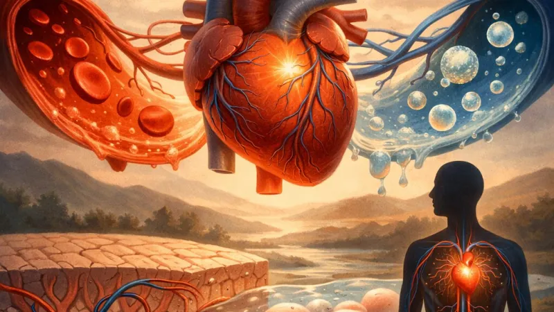
The human cardiovascular system is often compared to a grand network of roads, carrying precious cargo to every last cul-de-sac of our body. At the heart of this network, quite literally, lies an extraordinary organ: the heart itself, tirelessly pumping blood through an intricate arrangement of blood vessels. This essay will explore the structure and functions of the heart, arteries, capillaries, and veins, before examining the crucial role of tissue fluid in the maintenance of a healthy internal environment. Drawing upon both contemporary understanding and traditional knowledge cultivated in the United Kingdom’s academic and medical tradition, I will connect the dots between these components and show their interdependence in maintaining life.The Heart: Mainstream of Circulation
Situated in the thoracic cavity and roughly the size of a clenched fist, the human heart is built for efficiency. Structurally, it comprises four chambers – the right and left atria above, and the right and left ventricles below. This division separates oxygen-poor blood from oxygen-rich blood, ensuring that our tissues always receive adequately oxygenated supply. Heart valves – such as the tricuspid or bicuspid (mitral) valves – act as one-way gates, preventing the retrograde flow of blood.But beyond structure lies function: it acts as a muscular pump, with rhythmic contractions (the cardiac cycle) generating the very pressure that propels blood throughout the body’s highways: the blood vessels. The heart’s cycle splits into two distinct circulations. The pulmonary circuit channels blood from the right side through the lungs, where carbon dioxide is expelled and oxygen absorbed. The systemic circuit, by contrast, sends freshly oxygenated blood from the left side throughout the body, replenishing tissues and collecting waste. It is this relentless, coordinated effort which underpins our survival from one moment to the next.
Arteries: The Highways of Pressure
Having left the heart at considerable pressure, blood first travels into arteries. These vessels have adapted their structure to withstand and manage the surge produced by the heart’s contractions. Arteries possess thick walls layered with smooth muscle, elastic fibres, and collagen, rendering them both strong and flexible. The internal space, or lumen, is relatively narrow, which helps to maintain the high speed and pressure of blood flow.Elastic recoil is perhaps the most vital adaptation: for instance, the famous “dicrotic notch” seen in an arterial pressure graph reflects how the wall of the aorta rebounds after the heart’s pulse. This steadying effect ensures a smoother, more consistent blood flow to the body’s extremities. Additionally, the muscular component of arteries gives them the capacity for vasoconstriction and vasodilation, allowing regional control over blood distribution according to the body’s needs – for example, redistributing blood towards muscles during vigorous exercise, or shunting blood inward to conserve heat on a chilly London morning.
Another critical feature is the endothelium, a single-celled lining which provides an ultra-smooth surface, reducing resistance and deterring clot formation. British pathologists long ago identified damaged endothelium as a culprit in atherosclerosis, where fatty plaques lead to narrowing of arteries, restricting blood flow with serious consequences (as seen in classic cases of angina or myocardial infarction).
Capillaries: Fine Vessels of Exchange
Arteries soon divide into smaller arterioles and then the tiniest of vessels: capillaries. In this realm, structural economy is king – capillary walls comprise a single layer of flattened endothelial cells, sometimes only about 0.5 micrometres thick. Their diameter is just wide enough to allow red blood cells to squeeze through in single file, ensuring intimate proximity to surrounding tissue cells.This arrangement is no accident. Capillaries are the marketplace where goods are exchanged: oxygen and glucose diffuse out to nourish cells, while carbon dioxide and metabolic byproducts diffuse back in for removal. The diffusion process is driven by concentration gradients, and the ultra-thin walls facilitate rapid hampering-free exchange. The immense branching – think of the alveolar capillary network in the lungs or the close-packed vessels in the renal glomerulus – provides a colossal surface area to maximise exchange.
Variation exists depending on tissue requirements. For example, the blood-brain barrier is created by tight endothelial junctions, rigorously controlling passage for cerebral protection, whereas fenestrated capillaries in the kidney possess pores to allow extra filtration, essential for urine production.
Veins: The Low-Pressure Return Route
After traversing capillary beds, blood begins its homeward journey via the veins. In contrast with arteries, veins operate at much lower pressures and thus display a markedly different structure. Their walls are thinner, comprising less smooth muscle and fewer elastic fibres, as there is less need to accommodate pressure fluctuations. The lumen, however, is wider, enabling large volumes of blood to be stored or moved at once.Because blood flow is slower and pressure lower, veins face a risk of backflow, especially from areas below the heart. To combat this, veins contain pocket-like valves at intervals, similar in purpose to the locks of British canals such as those along the Grand Union. These venous valves ensure unidirectional flow back to the heart, aided by the contraction of surrounding skeletal muscles (“muscle pump”), and changes in pressure during breathing, especially prominent in the thoracic pump.
Veins also serve as a reservoir: in moments of need, such as sudden exertion or blood loss, the body can redirect blood from venous stores to maintain essential supplies. Their interplay with the lymphatic system is also vital, as the movement of fluid back from interstitial spaces often relies on both systems working in tandem.
The Creation and Function of Tissue Fluid
Between blood and cells lies tissue fluid, an often-overlooked yet crucial component of the internal environment. This clear, pale fluid bathes each cell, acting as an intermediary in the exchange of materials. Its formation owes itself to the balance of two primary forces across the capillary walls.Hydrostatic pressure, driven by the pumping heart, forces fluid out of the blood plasma at the arteriolar end of capillaries. This ultrafiltration process carries small solutes such as oxygen, glucose, amino acids, and ions into the interstitial space. However, not all plasma leaves: plasma proteins, too large to pass through capillary pores, remain inside and establish oncotic (osmotic) pressure. As blood traverses towards the venular end, falling hydrostatic pressure coupled with the osmotic draw of these proteins means water re-enters the capillaries, bringing some waste products with it.
Ideally, this cycle produces a dynamic balance. However, not all tissue fluid is reabsorbed; any surplus is collected by the lymphatic system, ultimately returning to the bloodstream via the thoracic duct near the left subclavian vein. When the balance tips due to illness or circulatory failure – as in heart failure or protein-losing conditions – tissue fluid accumulates, causing swelling or oedema, familiar to those who have seen patients with swollen ankles after prolonged standing.
Tissue fluid thus ensures cellular health, providing a medium for nutrient delivery, waste collection, and electrolyte balance. In the context of British healthcare, the management of oedema – whether pulmonary or peripheral – is a day-to-day clinical challenge, highlighting the importance of understanding these physiological mechanisms.
An Integrated Network: Collaboration Across the System
The heart, blood vessels, and tissue fluid work as a harmonious unit. The heart provides the driving force, arteries carry blood at high speed to capillaries, where vital exchanges occur, while veins complete the journey, returning blood for reoxygenation and waste removal. Throughout, homeostasis is maintained not by one structure but by the combined adaptation and regulation of all three.Regulation is key. The body finely tunes blood vessel diameter and cardiac output through neural (autonomic nervous system) and hormonal (such as adrenaline) controls, maintaining optimal blood pressure and tissue perfusion according to need – whether sprinting on the school field or writing quietly in a library.
Fluid balance, too, is crucial: even modest changes in tissue fluid can impact blood pressure, tissue health, and oxygen delivery, as any doctor or nurse in the NHS will attest. The adaptability of these systems explains our survival during variable activities and stresses, but also highlights how their failure can produce disease.
Conclusion
In sum, the cardiovascular system exemplifies the principle that structure serves function. Arteries, built for pressure; capillaries, built for exchange; veins, built for reservoir and return – all interwoven with tissue fluid, the silent mediator of cellular health. While the heart is often celebrated in literature and art as the seat of love and emotion, from Shakespeare’s verse to the poetry of T.S. Eliot, its true heroics lie in its ceaseless, mechanical precision, orchestrating an internal symphony of life. When this system falters, whether through structural disease or disruption of tissue fluid dynamics, health soon suffers – reminding us of the system’s pivotal importance.Additional Advice for Students
When revising this topic for A-levels or IB, do use clear, annotated diagrams to memorise vessel structure and location. Remember the functional significance of features like endothelium, valves, and lumen size. Relate your biological knowledge to real-life situations – such as noticing your pulse as an expression of arterial elasticity – to cement understanding. Consider the implications of disorders like atherosclerosis and oedema, both clinically relevant and ground for synoptic links in exams. Above all, focus on the language of the subject: terms like “ultrafiltration,” “vasoconstriction,” and “hydrostatic pressure” carry precise meanings that will set your answers apart.Embrace the elegance of the cardiovascular system: a marvel of living engineering, and an enduring subject of study in British medicine and education.
Rate:
Log in to rate the work.
Log in