Understanding the Musculoskeletal System: Anatomy and Function Explained
This work has been verified by our teacher: 2.04.2026 at 16:07
Homework type: Essay
Added: 1.04.2026 at 8:41
Summary:
Explore the musculoskeletal system’s anatomy and function to understand how muscles and bones work together for movement, support, and overall health.
The Musculoskeletal System: Foundations for Human Movement and Health
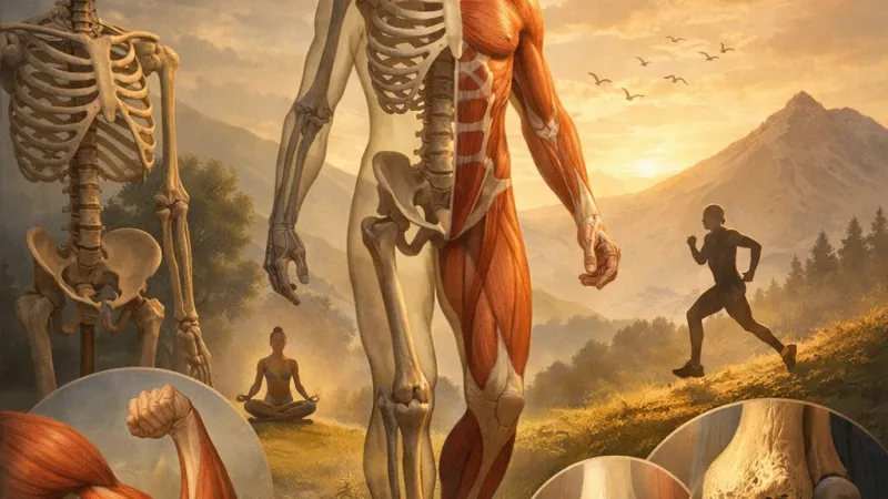
The musculoskeletal system underpins everything about our physical selves—facilitating movement, supporting stature, and protecting our most vital organs. It is a remarkable network comprised of bones, muscles, tendons, ligaments, cartilage, and connective tissues, each carrying out interdependent roles. Perhaps nowhere is this system's significance more evident than in our daily actions, whether ascending the steps of an old Victorian townhouse, rowing on the Thames, or simply standing in a queue. The collaborative action of these components allows such feats. In this essay, I propose to explore the anatomy and physiology of the musculoskeletal system, investigating the specific types of muscle tissue, the diverse functions of the skeleton, and how both collaborate. Central to this discussion will be a consideration of common disorders, exemplified by Marfan syndrome, which highlight the delicate interplay within the system. Mastery of this subject is indispensable for students in the health and biological sciences, serving as a foundation for more advanced understanding in clinical and therapeutic contexts.
---
Anatomy and Physiology of Muscle Tissue
General Characteristics of Muscle Tissue
Muscle tissue is unique in its fundamental property: the ability to contract. Contraction is the engine behind not just movement, but also posture, blood circulation, digestion, and heat generation. In classic British physiology, the sliding filament theory, first discerned using careful microscopy in the early twentieth century, describes how actin and myosin filaments within the muscle fibre slide past each other to produce contraction. This molecular ballet is powered by ATP and regulated by complex calcium ion dynamics. Consequently, muscle tissue is not a mere driver of movement; for instance, it plays a role in maintaining optimal body temperature, even contributing to the shivering response so familiar during a chilly winter’s morning in the Lake District.Classification of Muscle Tissue
Skeletal Muscle
Skeletal muscles are those associates with volitional movement—attached to bones by robust tendons. Their cellular structure is distinctive: elongated fibres, conspicuously striated under the microscope, and housing multiple nuclei per cell. Control is voluntary; it is the somatic nervous system that orchestrates a conductor’s gesture, a footballer’s kick, or the fine motor movements of a violinist. Functions range from generating power and movement to providing posture (as with the erector spinae muscles along the spine) and producing heat. Even at rest, muscles maintain a certain tension—muscle tone—crucial for stabilising joints, as observed in the reflexive and persistent contraction of postural muscles.Cardiac Muscle
Cardiac muscle is found only in the heart, an organ whose unrelenting contractions are poetically described by John Donne as “the clock of man.” Its cells are shorter than their skeletal counterparts, soft-branched, interconnected by intercalated discs—structures responsible for the rapid passage of electrical signals vital to coordinated heartbeat. Control here is involuntary: the sinoatrial node acts as the intrinsic pacemaker, whilst both nervous and hormonal signals can modulate rate and force, ensuring the heart adapts to rest or exertion. The heart’s specialisation is clear—where skeletal muscle rests, the heart must never tire.Smooth Muscle
Smooth muscle lines the walls of internal organs, blood vessels, and even the irises of the eyes—enabling peristaltic movements in the digestive tract, controlling the diameter of blood vessels, and modulating airflow and vision. These cells are spindle-shaped, lacking obvious striations, and contain a single nucleus. Their activity is involuntary, governed by the autonomic nervous system, hormones, and local chemical signals. Smooth muscle contractions are often coordinated en masse, thanks to gap junctions allowing ions and messages to spread cell-to-cell. This synchronicity is exemplified during childbirth, when waves of muscle contraction drive labour.---
The Skeletal System: Structure and Functional Roles
Components of the Skeletal System
The human skeleton consists of 206 bones at maturity, ranging from the diminutive ossicles of the inner ear to the long femur of the thigh. Bones are classified by their shapes—long (humerus), flat (sternum), short (carpals), and irregular (vertebrae). The architecture is complex, with dense compact bone forming the surface and light, spongy bone within, especially at the ends of longer bones. Cartilage, as found in the nose and at joints, provides flexible support, while tendons (muscle-to-bone connectors) and ligaments (bone-to-bone connections) confer stability. Synovial membranes line joints and secrete fluid to reduce friction, echoing Leonardo da Vinci’s musings on the body’s “living mechanics.”Primary Functions of the Skeletal System
Body Support
The skeleton is the framework around which the body is built. It must be rigid enough to support weight, especially in bipedal creatures like humans. Cartilage, though less rigid, provides the flexibility necessary in sites such as the ribcage—expanding and retracting with each breath.Protection of Vital Organs
Several bones in the skeleton serve chiefly as protectors: the skull encases the brain and sense organs, the vertebral column houses the spinal cord, the ribcage shields the heart and lungs from blows, and the pelvis encloses elements of the digestive and reproductive apparatus. The importance of such protection has been immortalised in literature—Shakespeare’s reference to “brains, born in a bone-box” underlines the sanctity of the skull.Enabling Body Movement
Bones alone are inert. Movement arises from the interplay of muscles and bones across joints. This biomechanical partnership allows for walking, writing, dancing, and every nuance between. Varieties of joints—hinge (elbow), ball-and-socket (shoulder, hip), and pivot (neck)—provide for different axes and degrees of movement. Ligaments and tendons are key to both permitting graceful motion and limiting excess, a duality exemplified by the anterior cruciate ligament of the knee, so often injured in sport.Mineral Storage and Haematopoiesis
Bones are more than architecture—they are dynamic reservoirs. They store minerals, predominantly calcium and phosphate, crucial for numerous physiological processes, including neural signalling and muscle contraction. Furthermore, the marrow within long bones is a cradle for haematopoiesis (the manufacture of blood cells), a process which, if disrupted, can have fatal consequences. Bone is constantly remodelled in response to stresses and metabolic requirements, a homeostatic process studied since the days of Sir William Hunter.---
Interactions Between Muscular and Skeletal Systems
The synergy between muscles and bones is at the heart of all movement. Muscles attach to bones via tendons, and when muscle fibres contract, the resultant force pulls on the bone, causing movement at the joint. This principle is demonstrated each time a person lifts an object, throws a cricket ball, or plays the piano. Agonist and antagonist muscle pairs (such as the biceps and triceps of the upper arm) allow for controlled and precise motion—while one contracts, the other relaxes, producing smooth action. Muscle tone, even at rest, is essential for maintaining posture and reducing the risk of falls or joint damage.Neural control is just as important. The somatic nervous system delivers instantaneous messages from the brain to muscles, while reflex arcs provide rapid, automatic responses to danger (such as withdrawing a hand from a hot kettle). Prolonged immobility, as might occur following injury or chronic illness, leads to complications: muscle atrophy and joint stiffness, limiting independence and quality of life.
---
Common Disorders and Clinical Implications
Overview of Musculoskeletal Disorders
Disorders affecting the musculoskeletal system are a significant source of pain, disability, and poor life quality in the UK. Arthritis, with its many forms, is among the most prevalent—osteoarthritis is common in the elderly, while rheumatoid arthritis can affect people of all ages. Muscular dystrophies, inherited conditions causing progressive muscle weakening, are recognised early in life and are the focus of much research at British institutions like the MRC Centre for Neuromuscular Diseases. Fractures, sprains, and repetitive strain injuries (like those affecting the wrists of musicians and typists) are also everyday realities.Genetic predisposition, occupational hazards, diet, and physical activity all play their parts. With an ageing population and modern sedentary lifestyles, burdens on NHS services continue rising, highlighting the urgency of prevention and innovation in treatment.
Marfan Syndrome: A Case Study
Genetic Basis and Pathophysiology
Marfan syndrome is an inherited disorder, passed through families in an autosomal dominant fashion. It stems from a mutation in the gene for fibrillin-1, an essential protein in connective tissue that imparts strength and elasticity. The result is weakened supporting structures, leaving the body susceptible to a variety of problems. While rare, the impact of this condition is profound.Clinical Manifestations
Marfan syndrome manifests in multiple systems. Skeletally, individuals may present with unusually long arms, legs, fingers—a condition called arachnodactyly—together with joint hypermobility, making even modest physical activity challenging. The most deadly features affect the heart and aorta: the risk of aortic aneurysm or rupture necessitates ongoing medical surveillance. Ocular complications are common—dislocation of the lens (ectopia lentis) and myopia impair vision, while some sufferers experience spontaneous lung collapse. The poet John Keats, though not diagnosed in his lifetime, is speculated by some historians to have had Marfan syndrome, given his tall, lean stature and early death from related complications.Diagnosis and Management
Diagnosis can be confirmed through genetic testing, together with clinical signs and imaging studies, such as echocardiography to assess the aorta. Management includes rigorous control of blood pressure, avoidance of contact sports, and, when necessary, surgical repair. Yet there is no cure. Support groups such as the Marfan Association UK provide vital education and advocacy, while multidisciplinary teams (doctors, geneticists, physiotherapists) guide care. Psychologically, living with a chronic, frequently misunderstood condition can be taxing, reinforcing the need for holistic management.---
Conclusion
The musculoskeletal system, from the cellular whispers of actin and myosin to the resilient scaffolding of bone, is a marvel of design and function. The variety within muscle tissue supports the specialised needs of our bodies—locomotion, blood flow, and digestion—while the bones provide a framework for living, an armature on which the wonders of human motion and protection hang. The close collaborations between muscles, bones, and nerves permit the level of control and adaptation that defines us as a species.Understanding disorders such as Marfan syndrome reveals the fragility of this system, and our imperative to protect and support it throughout life. Good nutrition, regular exercise, and prompt management of injuries or disease are not merely lifestyle choices—they are critical to preserving independence and overall wellbeing.
---
Rate:
Log in to rate the work.
Log in